Measuring aging with Physiological Cartography (Cartographie Physiologique®)
Among the many techniques used today to measure aging, physiological cartography allows to estimate a person’s physiological age. It was developed by the de Jaeger Institute, a private association founded by Dr. de Jaeger.
The functional aptitudes of the human body deteriorate with time. This can be associated with the aging process. As time goes by, the body finds it harder to compensate the micro-aggressions that life habits and the outside world put it through. Biological repair and regulation systems then become less efficient, and permanent biological perturbations appear. They characterize pathological states that are directly linked to aging. As these issues appear, they are joined by a progressive decrease of physical capabilities. Measuring the physiological age and the speed of aging can then be done by evaluating these biological and physical changes through time.
Physiological cartography is the sum of many biological and physical characteristcs that have a proven link to the aging process [1].


How Physiological Cartography can help you determine your physiological age and the speed at which you age
Patients are offered an array of tests that can inform them of the state of their general health, on which to base the estimates of their physiological health. In the end, patients will be told what to do in order to preserve their good health and life expectancy. Calibrating an “anti-aging” program can be done according to the needs of each patient and is built around four pillars: food, exercise, food supplements, and hormone therapy. The evolution of the patient’s health will be evaluated again a few years down the road, to follow up on the efficiency of the “anti-aging regimen” given by the medical team, so the “treatment” can be constantly adapted to each patient’s situation [2].

Physiological cartography consists in biological and physical tests to measure aging
These tests measure parameters with well known connections with aging processes in order to asses the longevity of the subjects. The values given by the different tests’ results are compared to reference tables that help precisely determine how advanced the person’s physiological age is, compared to their chronological age.
Here are some of the tests taken with the Physiological cartography method:
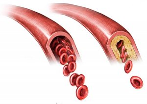
Assay for total and LDL (Low Density Lipoprotein) cholesterol: Cholesterol, a lipid, can be found in blood and is useful for many biological functions. When the body produces an excess of cholesterol, it can occasion sediments called atheromatous plaques. They can limit and even block the blood flow. Blood flow perturbation is responsible for many cardiovascular diseases and early deaths (ischemia, angina, aneurysm, atherosclerosis…) [3].
Determining body composition: Bio-impedance analysis and waist measuring are two good ways to identify a potential abdominal obesity, also called central obesity. Sub-cutaneous fat in the abdominal area as well as visceral fat help diffuse fatty acids into the blood stream through the portal venous system. Availability of free fatty acids favors an excessive mortality rate linked to metabolic diseases (insulin resistance, dyslimidemia…) which then lead to cardiovascular diseases, cancer, type 2 diabetes, etc. [4] [5]


Heart rate readings: A healthy person should have an average heart rate of 60 beats per minute. When it is too high, the heart rate can indicate poor physical condition, and become a liability on the long run, as a predictive factor of cardiovascular diseases, and sometimes even, of sudden death. A recent study showed that between two people with a 10bpm difference in heart rate, the one with the highest heart rate has a 9% increase in mortality risk. [6] [7]
Measure of the maximum oxygen consumption (VO2max): Maximum oxygen consumption is the maximum oxygen volume consumed during a physical effort, and it is measured in milliliter per minute. When a VO2max is around 40ml/min, it shows that the subject is in good physical condition, with great cardiovascular and respiratory systems. VO2max progressively decreases with age, which makes it interesting to measure [8] [9].
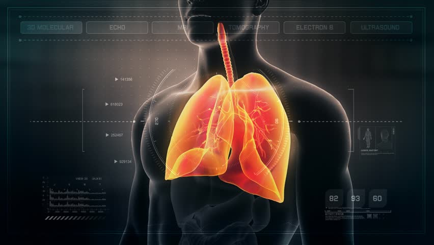
Measure of forced vital capacity (FVC): FVC represents the quantity of air we can breathe out after deeply breathing in, and shows the integrity of the respiratory system as a whole. Studies have shown that FVC tends to drop with age, some of them even offered mathematical models of the lung function representing the decline of the lungs due to age in healthy subjects [10] [11]. The test proved itself to be the best predictive test for life expectancy in the Framingham study [12].
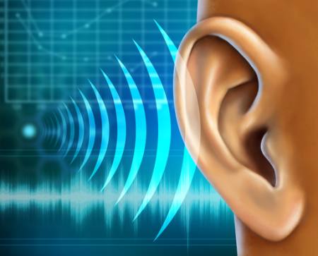
Hearing test: The human ear can hear sounds between 20 ans 20.000 hertz (HZ). Hearing loss happens with age, and is called age-related hearing loss. This loss is mostly due to the perpetual degradation of the ciliated cells in the internal ear, which are the cochlear sensory cells. They cannot regenerate. That is why a young subject can hear 18dB sounds when a 70 years old can only hear from 80dB[13] [14]. Research from 2014 indicates that NAD+ could play a role in the hearing loss process. It suggests that cochlear sensory cell decline would originate from a perturbation of NAD+ functions, and that preventive measures such as caloric restriction can be taken to compensate for hearing loss [15] [16].
Dosing auto-antibodies: Antibodies are protein complexes in the immune system. They defend the body against potential pathogens or foreign objects. It is only logical that auto-antibodies are molecules that the body deploys as a defense against itself; this results in tissue damage and chronic inflammation. Auto-immunity is a malfunction of the immune system when the body starts to fight against itself. When the auto-antibodies count is high in plasma, it shows a pathological state. However, with age, the levels of auto-antibodies in plasma increases without necessarily meaning that there is a hidden disease in the body [17] [18]. The phenomenon could be caused by aged immunity cell lines (lymphocytes, plasmocytes…) that would entail perturbations and destroy the balance of the immune system.
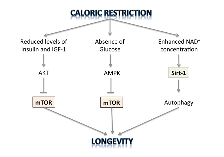
Measuring blood sugar levels: Knowing the levels of blood sugar can help diagnose some metabolic diseases (type 1 and 2 diabetes, gestational diabetes, “SMeT”), pancreatic diseases (pancreatitis, cystic fibrosis, haemochromatosis…) or diverse genetic diseases (Down’s syndrome, Prader-Willy syndrome…) Nevertheless, beyond giving us information on any pathological state, measuring blood sugar is a good indicator of the body’s state of caloric restriction, which was proven to be beneficial for longevity [19] [20] [21]. Hyperglycemia can negatively affect the structural integrity of proteins and DNA [22] [23].
Unipodal stance test: This consists in standing on one leg, eyes closed. It helps to monitor the integrity of the inner ear, of the central and peripheral neurological systems, and the muscular and osteotendinous states (sarcopenia). The older the person is, the harder it will be to stay balanced on one leg [24].
Measuring aging with the physiological cartography: pros and cons
Physiological cartography allows to estimate the physiological age and speed of aging of the patient by monitoring them for several years.
Aging is, in fact, specific to each person. That is why in order to appreciate the true organic status of a person, as well as their physiological age, there is no other way but to measure their aging at different times to see how it evolves.
The criteria and markets considered in this technique are linked to the risk to develop diseases that were proven to be deadly. Our different organs don’t age at the same speed, and the tests taken with this method allow to check for a wide range of biological and physical functions. For all of these reasons, this is a well-rounded method to measure aging.
Furthermore, some of the tests, such as forced vital capacity or auto-antibodies levels are very interesting since the studies showing age disturbs their functions are based on cohorts of healthy subjects of diverse age ranges. Such markers deserve to be used more often to measure a “healthy aging”. In order to determine the real biological age of a person, one needs to see beyond the pathological criteria.
Where ?
The physiological cartography method to measure aging is a brand that belongs to Dr. de Jaeger. Today, it is only available at the de Jaeger Institute in Paris, which he founded himself.
All our articles on the Metrology of Aging
Measuring aging: what is your biological age?

Measuring our biological age and take consistent dispositions to avoid it, it’s a dream come true !
Part 1: Measuring aging: Cartographie PhysiologiqueⓇ
 This method to measure aging and physiological age measures the many physical and biological characteristics that were definitely linked to the aging process (cholesterol levels, unipedal stance test, central obesity…)
Part 2: Measuring aging: the eighteen markers of Duke University

This method follows the decline of many organic systems (cardiovascular, lungs, kidneys, liver, teeth, and immune system) by quantifying 18 markers for chronic diseases linked to aging. This technique was developed by a research team from Duke University, under the supervision of Dr. Daniel Belsky.
Part 3: DNA methylation, a promising lead towards the measure of aging?
 DNA methylation levels grow with the number of divisions a cell undergoes. The number of cell divisions grows with time. Therefore, measuring the methylation of DNA helps to quantify aging. Treating DNA with bisulfites allows to convert non-methylated cytosines into uraciles, so the methylated cytosines can easily be identified. We deduce a physiological age from their numbers.
Part 4: Telomeres, at witnesses of the physiological age, as a tool to measure aging
 Telomeres can be found at the end of our chromosomes, and get shorter with age. There are many techniques to measure their length (TRFs, Q-Fish, qPCR, STELA…) based on the Southern Blot principle, PCR and in situ hybridization.
Farah Bahou

Author
Auteure
Farah studied biochemistry, therapeutics and molecular and biopharmaceutical innovation at Aix-Marseille university and Paris 7 Diderot university.
More about the Long Long Life team
Farah a étudié la biochimie, la thérapeutique et les innovations moléculaires et biopharmaceutiques à l’université d’Aix-Marseille et à l’université Paris 7 Diderot.
En savoir plus sur l’équipe de Long Long Life
Dr Guilhem Velvé Casquillas

Author/Reviewer
Auteur/Relecteur
Physics PhD, CEO NBIC Valley, CEO Long Long Life, CEO Elvesys Microfluidic Innovation Center
More about the Long Long Life team
Docteur en physique, CEO NBIC Valley, CEO Long Long Life, CEO Elvesys Microfluidic Innovation Center
En savoir plus sur l’équipe de Long Long Life
[1] http://www.institutdejaeger.com/
[2] La Nouvelle Méthode anti-âge (Odile Jacob, 2008), Christophe de Jaeger
[3] Prospective Studies Collaboration. (2007). Blood cholesterol and vascular mortality by age, sex, and blood pressure: a meta-analysis of individual data from 61 prospective studies with 55 000 vascular deaths. The Lancet, 370(9602), 1829-1839.
[4] Carmienke, S., Freitag, M. H., Pischon, T., Schlattmann, P., Fankhaenel, T., Goebel, H., & Gensichen, J. (2013). General and abdominal obesity parameters and their combination in relation to mortality: a systematic review and meta-regression analysis. European journal of clinical nutrition, 67(6), 573-585.
[5] Hocking, S., Samocha-Bonet, D., Milner, K. L., Greenfield, J. R., & Chisholm, D. J. (2013). Adiposity and insulin resistance in humans: the role of the different tissue and cellular lipid depots. Endocrine reviews, 34(4), 463-500.
[6] Zhang, D., Shen, X., & Qi, X. (2015). Resting heart rate and all-cause and cardiovascular mortality in the general population: a meta-analysis. Canadian Medical Association Journal, cmaj-150535.
[7] Zhao, Q., Li, H., Wang, A., Guo, J., Yu, J., Luo, Y., … & Guo, X. (2017). Cumulative Resting Heart Rate Exposure and Risk of All-Cause Mortality: Results from the Kailuan Cohort Study. Scientific Reports, 7, 40212.
[8] Betik, A. C., & Hepple, R. T. (2008). Determinants of VO2 max decline with aging: an integrated perspective. Applied physiology, nutrition, and metabolism, 33(1), 130-140.
[9] DeLorey, D. S., Kowalchuk, J. M., & Paterson, D. H. (2004). Effects of prior heavy-intensity exercise on pulmonary O2 uptake and muscle deoxygenation kinetics in young and older adult humans. Journal of Applied Physiology, 97(3), 998-1005.
[10] Falaschetti, E., Laiho, J., Primatesta, P., & Purdon, S. (2004). Prediction equations for normal and low lung function from the Health Survey for England. European Respiratory Journal, 23(3), 456-463.
[11] Hankinson, J. L., Odencrantz, J. R., & Fedan, K. B. (1999). Spirometric reference values from a sample of the general US population. American journal of respiratory and critical care medicine, 159(1), 179-187.
[12] https://www.framinghamheartstudy.org/fhs-bibliography/index.php
[13] Bainbridge, K. E., & Wallhagen, M. I. (2014). Hearing loss in an aging American population: extent, impact, and management. Annual review of public health, 35, 139-152.
[14] Someya, S., Yamasoba, T., Weindruch, R., Prolla, T. A., & Tanokura, M. (2007). Caloric restriction suppresses apoptotic cell death in the mammalian cochlea and leads to prevention of presbycusis. Neurobiology of aging, 28(10), 1613-1622.
[15] Fujimoto, C., & Yamasoba, T. (2014). Oxidative stresses and mitochondrial dysfunction in age-related hearing loss. Oxidative medicine and cellular longevity, 2014.
[16] Kim, H. J., Oh, G. S., Choe, S. K., Kwak, T. H., Park, R., & So, H. S. (2014). NAD+ metabolism in age-related hearing loss. Aging and disease, 5(2), 150.
[17] Nagele, E. P., Han, M., Acharya, N. K., DeMarshall, C., Kosciuk, M. C., & Nagele, R. G. (2013). Natural IgG autoantibodies are abundant and ubiquitous in human sera, and their number is influenced by age, gender, and disease. PLoS One, 8(4), e60726.
[18] Higashino, A., Kageyama, T., Kantha, S. S., & Terao, K. (2011). Detection of elevated antibody against calreticulin by ELISA in aged cynomolgus monkey plasma. Zoological science, 28(2), 85-89.
[19] Speakman, J. R., & Mitchell, S. E. (2011). Caloric restriction. Molecular aspects of medicine, 32(3), 159-221.
[20] Ravussin, E., Redman, L. M., Rochon, J., Das, S. K., Fontana, L., Kraus, W. E., … & Smith, S. R. (2015). A 2-year randomized controlled trial of human caloric restriction: feasibility and effects on predictors of health span and longevity. The Journals of Gerontology Series A: Biological Sciences and Medical Sciences, 70(9), 1097-1104.
[21] Schlotterer, A., Kukudov, G., Bozorgmehr, F., Hutter, H., Du, X., Oikonomou, D., … & Sayed, A. (2009). C. elegans as model for the study of high glucose–mediated life span reduction. Diabetes, 58(11), 2450-2456.
[22] Haus, J. M., Carrithers, J. A., Trappe, S. W., & Trappe, T. A. (2007). Collagen, cross-linking, and advanced glycation end products in aging human skeletal muscle. Journal of applied physiology, 103(6), 2068-2076.
[23] Pinkas, A., & Aschner, M. (2016). Advanced glycation end-products and their receptors: related pathologies, recent therapeutic strategies, and a potential model for future neurodegeneration studies. Chemical research in toxicology, 29(5), 707-714.
[24] Roemer, K., & Raisbeck, L. (2015). Temporal dependency of sway during single leg stance changes with age. Clinical Biomechanics, 30(1), 66-70.

